This podcast episode is worth 0.27 CPD hours. Upgrade to Pro
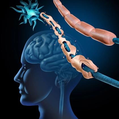
In this episode, Dr Roger Henderson explores multiple sclerosis (MS), a chronic immune-mediated disorder of the central nervous system (CNS) that can present in a remarkably diverse range of ways. For GPs, recognising MS is not simply about identifying an abnormal magnetic resonance imaging (MRI) scan. It is about understanding the clinical pattern, localising neurological symptoms, recognising dissemination in space and time, and considering the important alternative diagnoses that can mimic the disease. The episode looks at the underlying immunology and pathology, including the roles of inflammation, demyelination and neuroaxonal injury, before moving on to the clinical presentations and diagnostic approach. It also discusses current principles of disease-modifying treatment, relapse management, rehabilitation and symptom control, while considering the often substantial cognitive, psychological and functional burden of MS. Most importantly, it focuses on practical lessons for primary care: recognise the syndrome, interpret investigations in context and treat the patient, not simply the scan.
Key take-home points
- MS is a chronic immune-mediated disease of the CNS characterised by inflammation, demyelination, gliosis and neuroaxonal injury.
- Most patients initially have relapsing-remitting MS, with neurological symptoms developing over hours to days and lasting at least 24 hours. Recovery may be complete or incomplete, with some patients subsequently developing progressive disability.
- Primary progressive MS accounts for a smaller proportion of cases and is characterised by gradual accumulation of disability from disease onset without a preceding relapsing-remitting phase.
- Think anatomically when assessing suspected MS: optic nerves, brainstem, spinal cord, cerebellum and cerebral hemispheres can all be involved.
- Optic neuritis is a classic presentation, typically causing subacute monocular visual loss, often with pain on eye movement.
- Spinal cord involvement can produce a wide range of symptoms, including weakness, sensory disturbance, sphincter dysfunction and Lhermitte phenomenon.
- The non-motor burden of MS is substantial. Fatigue, cognitive impairment, mood disturbance, sleep problems, bladder and bowel dysfunction, and sexual dysfunction can have a major impact on quality of life.
- MS has a multifactorial aetiology, involving genetic susceptibility, immune dysregulation and environmental factors, with Epstein–Barr virus now recognised as an important epidemiological association.
- Pathology reflects both inflammation and neurodegeneration. Demyelination and axonal injury can continue to contribute to disability even when overt relapses become less frequent.
- The diagnosis of MS depends on the overall clinical picture rather than a single test. MRI is central, with cerebrospinal fluid (CSF) oligoclonal bands and selected blood tests providing additional diagnostic information where appropriate.
- Remember dissemination in space and dissemination in time. Lesions should demonstrate involvement of characteristic CNS regions, while evidence of disease occurring at different points in time can support the diagnosis; contemporary diagnostic criteria also allow CSF and other biomarkers to contribute in appropriate circumstances.
- An abnormal MRI does not automatically mean MS. The imaging abnormalities must fit the clinical syndrome, and important alternative diagnoses need to be considered.
- Red flags should prompt reconsideration of the diagnosis, including symptoms reaching maximum severity within minutes, encephalopathy, prominent systemic features, multiple cranial neuropathies, progressive ataxia or rapidly progressive disease.
- Treating MS involves more than preventing relapses. Disease-modifying therapy aims to reduce inflammatory disease activity and disability accumulation, while rehabilitation and symptomatic treatment address the patient's day-to-day functional problems.
- For GPs, the key message is to manage the patient rather than simply the scan: localise the syndrome, establish its temporal profile, interpret MRI findings in clinical context, exclude important mimics and address the physical, cognitive and psychological consequences of the disease.
Key references
- NICE. 2022. https://www.nice.org.uk/guidance/ng220.
- Thompson AJ, et al. Lancet Neurol. 2018;17(2):162-173. doi: 10.1016/S1474-4422(17)30470-2.
- Wattjes MP, et al. Lancet Neurol. 2021;20(8):653-670. doi: 10.1016/S1474-4422(21)00095-8.
- Montalban X, et al. Mult Scler. 2018;24(2):96-120. doi: 10.1177/1352458517751049.
- Solari A, et al. Eur J Neurol. 2020;27(8):1510-1529. doi: 10.1111/ene.14248.
Create an account to add page annotations
Annotations allow you to add information to this page that would be handy to have on hand during a consultation. E.g. a website or number. This information will always show when you visit this page.