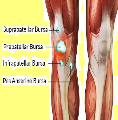
Pes anserine bursitis
Anserine Bursitis
The anserine bursa is located inferiorly to the knee joint on the medial aspect of the upper calf beneath the conjoined tendons of sartorius, gracilis and semi-tendinosis at the level of the tibial tuberosity
- Pes anserine bursitis is a clinical entity that is associated with pain at the medial knee and upper tibial region
- term 'pes anserine' comes from the Latin referring to "goose's foot," which the tendinous structures of the semitendinosus, gracilis, and sartorius muscles are said to resemble as they join to insert at the medial knee
- a more generic term, pes anserine pain syndrome, has been applied to refer to medial knee pain which may or may not include inflammation of the bursa sac
Aetiology:
- mechanical derangement, direct trauma, obesity, and overuse have all been implicated in the development of pes anserine bursitis - inflammation of the pes anserine bursae may not be the primary pathology but rather a sequela of earlier knee complications (1)
- medial knee osteoarthritis is an early and common finding in patients with this condition - association of pes anserine pain (but not necessarily bursitis) with concomitant osteoarthritis was noted in one study to be over 90% (2)
- some sports may make increase the risk of development of pes anserine inflammatory conditions including running, basketball, and racquet sports
Clinical features:
- when present, patients may complain of an aching pain on the medial aspect of the knee and localized tenderness to palpation
- in particular with rising from a seated position, going upstairs, or sitting with their legs crossed.
- may be subjective complaints of muscle weakness and decreased range of motion of the knee joint
- on examination
- tenderness is invariably present over at the insertion of the pes anserine tendons ("goose's foot") at the medial knee and upper medial tibia - tenderness will likely be present in the medial knee joint and may extend along the proximal, medial tibial region
- swelling is a variable feature
- with knee flexion to 90 degrees
- tenderness may be palpated along the medial tendinous structures of the pes anserine group as they travel to insert along the medial tibial region
- pes anserine bursa lies directly beneath the tendons at their insertion
Investigation/Assessment:
In general imaging does not assist in diagnosis of anserine bursitis. Investigations that may be undertaken:
- plain knee radiographs - obtained to observe for any underlying bony abnormalities including osteoarthritis
- ultrasound - used as an adjunct to evaluate for other causes of localized swelling, including joint effusions.
- MRI - may be useful in the assessing for knee pathology and ruling out alternative diagnoses.
Treatment
- generally supportive with steroid injections reserved for refractory cases.
Bursae of the knee:

Bursitis of the knee video - different types of bursitis
Reference:
- Pompan DC. Pes Anserine Bursitis: An Underdiagnosed Cause of Knee Pain in Overweight Women. Am Fam Physician. 2016 Feb 01;93(3):170.
- Alvarez-Nemegyei J et al. Prevalence of rheumatic regional pain syndromes in Latin-American indigenous groups: a census study based on COPCORD methodology and syndrome-specific diagnostic criteria. Clin. Rheumatol. 2016 Jul;35 Suppl 1:63-70.
Related pages
Create an account to add page annotations
Annotations allow you to add information to this page that would be handy to have on hand during a consultation. E.g. a website or number. This information will always show when you visit this page.