Echocardiography uses ultrasound to provide a real-time image of the heart. Notably:
- it is non-invasive and is thought to be harmless
- the ultrasound generator and receiver is a piezo- electric crystal
- for high resolution images a high frequency ultrasound must be used - this is 2 MHz for adults and 7 MHz for infants
Notes (1):
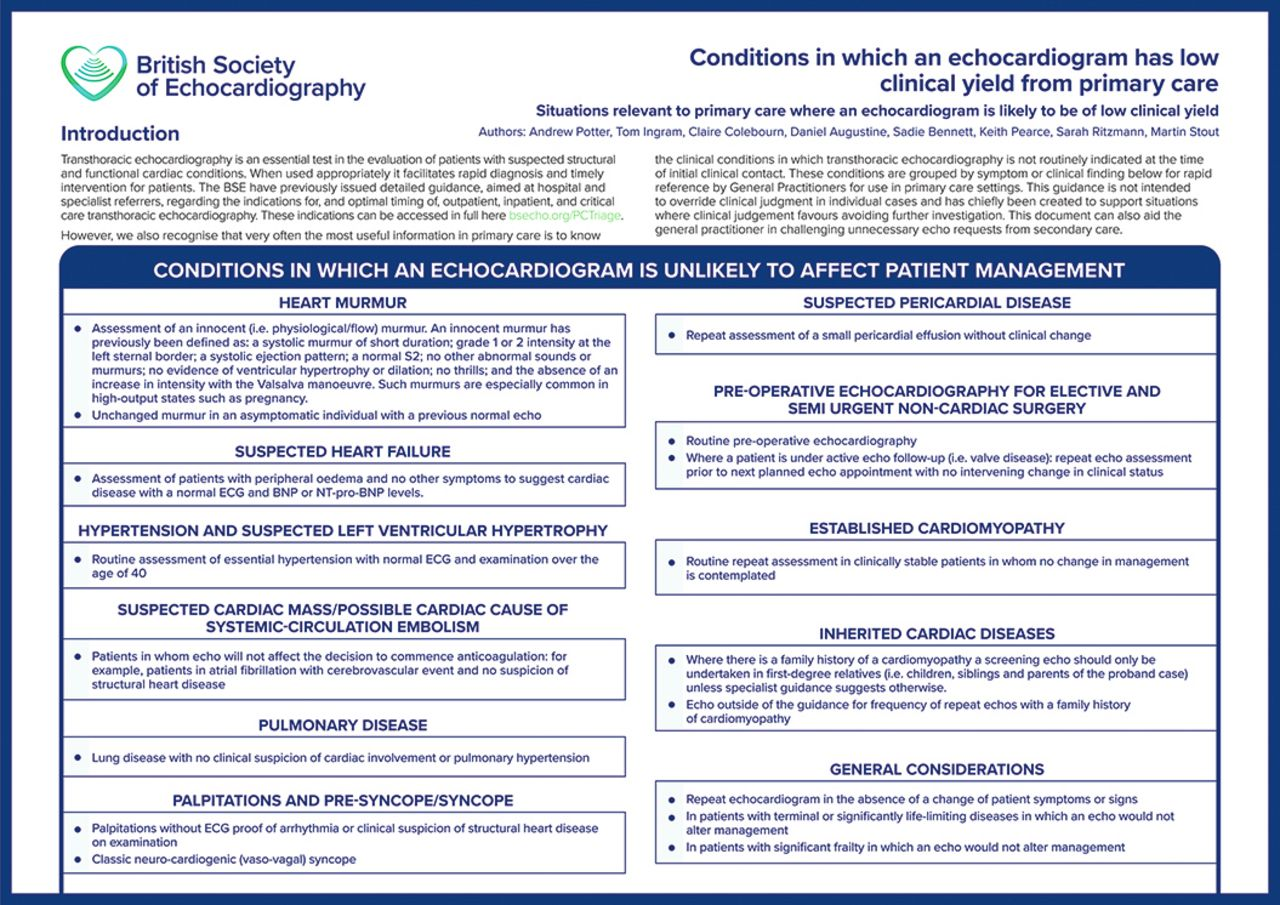
- heart failure:
- in the absence of a significant cardiac history, and with a normal physical examination and 12-lead electrocardiogram (ECG), then the presence of simple cardiomegaly (without pulmonary congestion or other findings suggestive of cardiac disease) on a chest X-ray does not warrant an echocardiogram as the likely yield of identifying significant cardiac pathology is low
- hypertension:
- an echocardiogram is not indicated routinely to evaluate patients with hypertension, particularly when a normal 12-lead ECG and physical examination is present (1)
Reference:
Related pages
Create an account to add page annotations
Add information to this page that would be handy to have on hand during a consultation, such as a web address or phone number. This information will always be displayed when you visit this page