Pulmonary embolus (PE) occurs when a clot from a vein, originating in the venous sinuses of the calf or the femoral vein or the pelvis, detaches and becomes lodged in the pulmonary arterial tree (1).
Occasionally the right side of the heart is a source of a pulmonary embolus.
Deep vein thrombosis (DVT) is the formation of blood clots in deep veins of the legs (1). In a majority of patients, PE is a consequence of DVT (2)
- when sensitive diagnostic methods were used, DVT was detected in around 70% of patients with PE (2)
- clinically important PEs originate from proximal DVT of the leg e.g. - popliteal, femoral or iliac veins (3)
Venous thromboembolism (VTE) is the term used to describe a thrombus in a vein which may detach from the site of origin and travel through blood to a distant site, a phenomenon called embolism (1). PE and DVT represent different clinical manifestations of VTE (2).
An estimated 12-36% of patients with PE are misdiagnosed during initial evaluation in emergency departments or primary care clinic (4)
Non thrombotic pulmonary emboli are rare. Causes include:
- septic emboli
- fat emboli
- amniotic fluid
- venous air embolism
- intravascular foreign bodies
- tumor emboli (2)
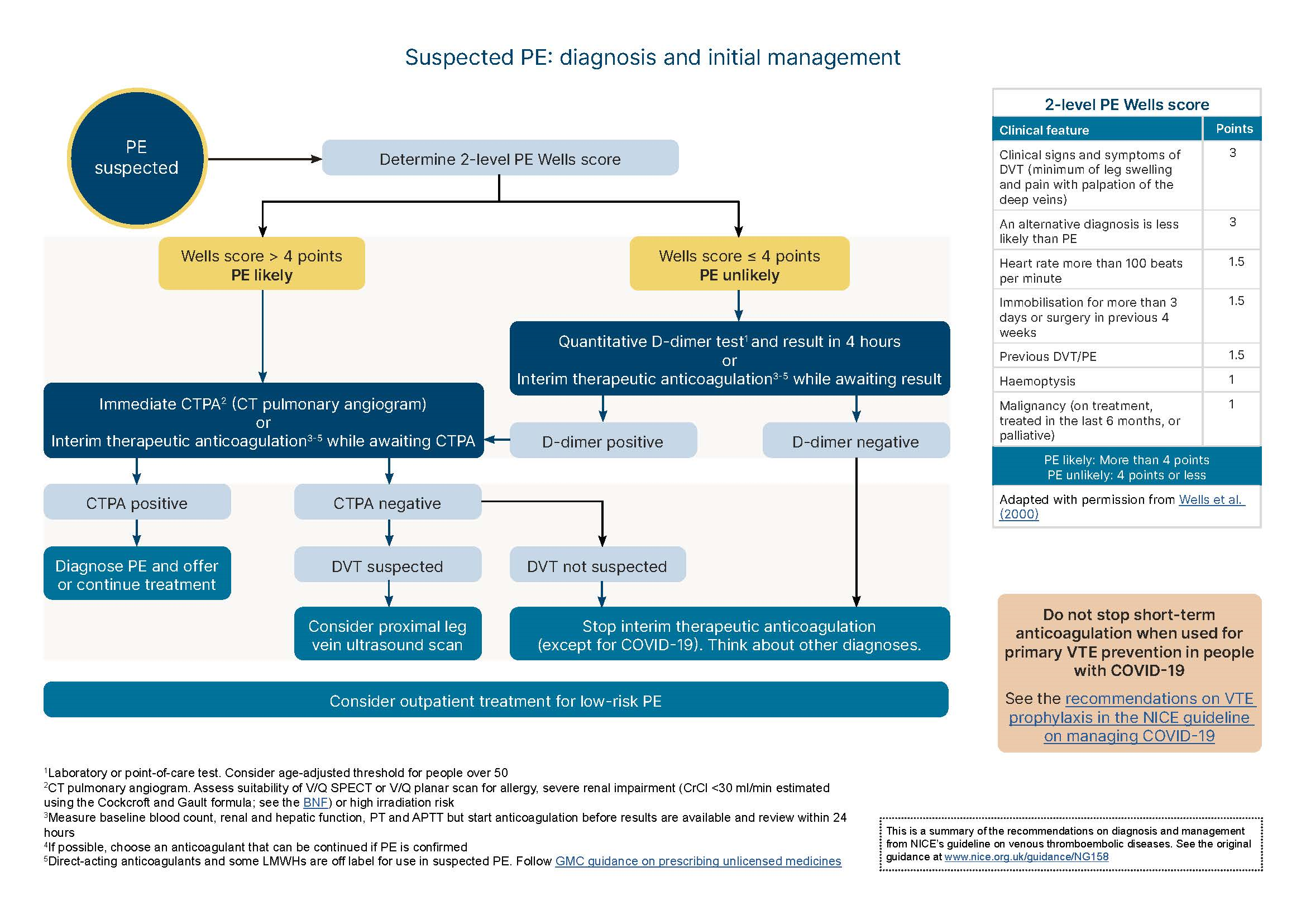
Computed tomography pulmonary angiogram (CTPA) scan
- most common diagnostic imaging modality for PE
- V/Q SPECT (ventilation-perfusion single photon emission CT) is a scintigraphic modality that captures three dimensional images and offers equal diagnostic accuracy as CTPA while delivering a lower absorbed dose of radiation (4)
Reference:
- (1) National Clinical Guideline Centre (NICE) 2010. Venous thromboembolism: reducing the risk of venous thromboembolism (deep vein thrombosis and pulmonary embolism) in patients admitted to hospital.
- (2) Torbicki A et al. Guidelines on the diagnosis and management of acute pulmonary embolism: the Task Force for the Diagnosis and Management of Acute Pulmonary Embolism of the European Society of Cardiology (ESC). Eur Heart J. 2008 ;29(18):2276-315.
- (3) Ramzi DW, Leeper KV. DVT and pulmonary embolism: Part I. Diagnosis. Am Fam Physician. 2004;69(12):2829-36.
- (4)NICE (August 2023). Venous thromboembolic diseases: diagnosis, management and thrombophilia testing
- (5) Maughan B C, Jarman A F, Redmond A, Geersing G, Kline J A. Pulmonary embolism BMJ 2024; 384 :e071662 doi:10.1136/bmj-2022-071662
Related pages
Create an account to add page annotations
Add information to this page that would be handy to have on hand during a consultation, such as a web address or phone number. This information will always be displayed when you visit this page